Veterinary radiology is often used as a synonym for radiographs, and for most of its history that was fair. It is no longer. As CT has spread into general and emergency practice, the clinically important question has shifted from can we image this patient to which imaging answers this question, and what does a specialist see in the result. Below are seven specific things a CT scan reveals that a radiographic study, taken well, frequently cannot.
By the RadsForVets editorial team, with clinical direction from Miriam M. Shanaman, VMD, MS, DACVR, Chief Medical Officer and board-certified veterinary radiologist. Published August 21, 2026.
Key takeaways
- Radiographs superimpose anatomy. CT removes superimposition, which is why equivocal skull, thorax and pelvis studies so often resolve on cross-section.
- Pulmonary nodules several millimetres across are routinely invisible on radiographs and routinely visible on CT, which changes surgical staging decisions.
- One CT acquisition can triage several body regions in a trauma patient in less time than a full radiographic series takes to position.
- A CT study contains hundreds of images across multiple windows; interpretation time and training scale with that, not with patient size.
- Contrast phase timing is often the difference between seeing a vascular anomaly and missing it entirely.
- RadsForVets reads CT, including contrast-enhanced and multi-phase studies, at the same STAT target of about one hour.
Why cross-section changes the answer
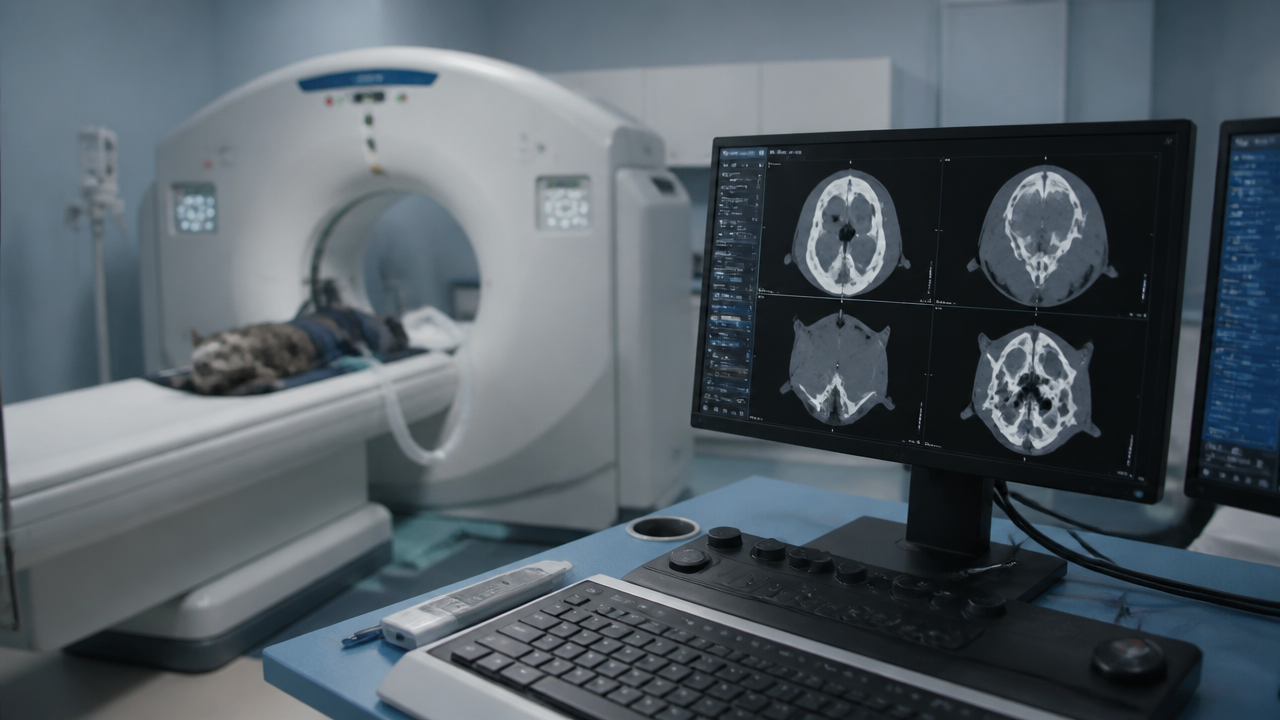
A radiograph is a shadow. Every structure between the tube and the detector contributes to one flattened image, so overlapping anatomy either hides pathology or creates it. CT acquires volumetric data and reconstructs it as thin slices viewable in any plane, at a contrast resolution radiography cannot approach for soft tissue.
Worth saying plainly: RadsForVets is rebuilding its search presence from a standing start and none of our tracked terms currently rank in the top 100. Nothing in this article is shaped around protecting a position. It is written the way a radiologist would explain the modality to a referring colleague. Our pillar guide to veterinary CT scan interpretation covers protocols and reporting in more depth.
1. Occult and complex fractures
The classic scenario is a patient that is clearly painful and lame with radiographs read as normal or equivocal. Non-displaced fracture lines, incomplete fissures, and fractures in anatomically busy regions such as the pelvis, tarsus, carpus and skull disappear into superimposed bone on projection imaging.
Where CT changes management
- Pelvic fractures, where the number and displacement of fracture sites determines whether surgery is needed at all.
- Carpal and tarsal injuries, where small avulsions dictate whether arthrodesis is on the table.
- Articular fracture involvement, which is frequently underestimated on radiographs and directly changes prognosis.
- Pre-surgical planning, because a surgeon can rotate the reconstruction and see the fragment configuration before opening.
2. Lung nodules below radiographic resolution
This is the most consequential difference in oncology work. Detecting a pulmonary nodule on thoracic radiographs generally requires it to be several millimetres across and favourably positioned against the surrounding lung. CT detects considerably smaller nodules and detects them in locations radiographs obscure, such as adjacent to the heart or the diaphragm.
| Thoracic radiographs | Thoracic CT | |
|---|---|---|
| Smallest reliably detected nodule | Larger nodules only | Substantially smaller nodules |
| Superimposition | Heart, diaphragm and ribs obscure regions | None; each slice is independent |
| Acquisition | Three views, awake in many patients | Seconds, sedation or anaesthesia usual |
| Typical use | Screening, follow-up, unstable patients | Staging before surgery, equivocal radiographs |
The decision this drives is concrete: whether to proceed with an amputation or major resection in a patient whose radiographs looked clean.
3. Nasal, dental and middle ear disease
The skull is the region where radiographs are weakest, because the anatomy is dense, symmetrical and heavily superimposed. Chronic nasal discharge, chronic otitis, and dental disease with suspected extension are the three presentations that most reliably justify CT.
- Nasal cavity. Turbinate destruction, fluid versus soft tissue, unilateral versus bilateral involvement, and critically the integrity of the cribriform plate before treatment planning.
- Tympanic bullae. Fluid, soft tissue and bulla wall thickening, all of which are essentially unassessable on radiographs in many patients.
- Dental. Tooth root involvement, bone lysis and the extent of any mass beyond what intraoral radiography can display.
4. Abdominal masses, vascular involvement and portosystemic shunts
Ultrasound remains excellent for abdominal parenchyma and is often the right first modality. Where CT adds decisively is in defining a mass in relation to its blood supply and the structures around it, which is the surgeon's actual question.
- Organ of origin, when a large mass has displaced everything around it and ultrasound cannot establish where it began.
- Vascular invasion or encasement, which frequently determines resectability.
- Portosystemic shunt anatomy, where multi-phase contrast CT angiography maps the anomalous vessel precisely enough to plan attenuation.
- Peritoneal and lymph node staging in a single acquisition alongside the thorax.
Contrast timing is doing most of the work in these studies. A vascular anomaly imaged in the wrong phase can be invisible, which is one reason protocol advice before the scan is part of the radiologist's job rather than an afterthought.
5. Acute trauma triage across multiple regions in one scan
A patient hit by a car may have thoracic, abdominal, pelvic and spinal injuries at once. Radiographing all four regions well means repeated positioning of a painful, unstable animal, and each region is interpreted in isolation.
A single CT acquisition covers them together in seconds of scan time and produces one dataset that can be reviewed region by region. For emergency practice the change is not only diagnostic yield but patient handling: less repositioning, shorter time off monitoring, and a prioritised problem list back quickly. Coupled with a one hour STAT read, the practical effect is that the surgical decision happens the same evening. That workflow effect is the same one described in what veterinary radiology services actually include.
6. What a veterinary CT scan actually shows
Stepping back from specific diseases, it is worth stating what a veterinary CT scan shows in general terms, because this is the point most owner conversations and many clinical decisions turn on.
| Tissue or structure | What CT resolves | Clinical relevance |
|---|---|---|
| Bone | Cortical and trabecular detail, fine fracture lines, lysis | Fracture configuration, tumour margins, surgical planning |
| Lung | Parenchymal detail slice by slice | Small nodules, patterns, bullae, contusion extent |
| Soft tissue masses | Margins, internal structure, enhancement pattern | Resectability and biopsy targeting |
| Vessels | Contrast-filled anatomy in timed phases | Shunts, invasion, vascular supply to a mass |
| Air-filled spaces | Nasal cavity, sinuses, bullae | Chronic nasal and middle ear disease |
| Brain and spinal cord | Limited compared with MRI | CT is a screening tool here; MRI is preferred |
The last row matters as much as the others. CT is not a universal upgrade. For brain and spinal cord parenchyma, MRI remains the better modality, and part of the value of asking a radiologist before scanning is being told which machine answers your question.
7. Why specialist interpretation of CT specifically matters
A three-view radiographic study is three images. A CT study is commonly several hundred, and with multi-phase contrast can be well over a thousand, each requiring review at more than one window setting because bone, soft tissue and lung are read differently from the same data.
- Volume. Systematic review of that many images is a trained discipline, not a matter of scrolling faster.
- Cross-sectional anatomy. Recognising normal in a transverse plane, across species and breeds, is a separate skill from reading projections, and it is what residency spends years building.
- Windowing. A pulmonary nodule invisible at soft tissue window is obvious at lung window. Findings are missed at the display level before they are ever missed at the diagnostic level.
- Reformatting. Reconstructing in sagittal, dorsal or oblique planes is often what converts an ambiguous finding into a definite one.
- Incidentals. Higher sensitivity surfaces findings unrelated to the presenting problem. Deciding which of those warrant action is judgment, and over-calling them causes real harm.
That combination is why access to the scanner and access to the reader are two different problems. A practice can solve the second today. See what a veterinary radiologist is for the credential behind that reading, the RadsForVets overview for turnaround tiers and modality coverage, or contact our team to send a study.
Free for veterinary teams
CT submission and protocol quick sheet
A practical reference for practices new to sending CT studies out for interpretation, written by radiologists who read them daily.
- Which clinical questions justify CT over radiographs or ultrasound
- Contrast phase selection for vascular and mass evaluation
- What to include in the submission so the report answers your question
No contracts. No minimums. Radiologists on shift every day of the year.
Frequently asked questions
This set leans more heavily on reasoned clinical questions than our other two articles, because far fewer tracked AI-search prompts exist specifically for veterinary CT than for radiology services or the radiologist credential. The first group comes largely from real search autocomplete data; the later groups reflect what practices ask us directly.
What CT shows
Why would a dog need a CT scan?
Most commonly when radiographs are equivocal and the answer changes management: suspected occult fracture, chronic nasal disease, middle ear disease, lung metastasis screening before surgery, complex abdominal masses, portosystemic shunt evaluation, and multi-region trauma. CT is also used to plan surgery, because a surgeon can see relationships between structures that radiographs superimpose.
What does a CT scan show in dogs?
Cross-sectional anatomy at sub-millimetre resolution with no superimposition: bone detail including non-displaced fracture lines, lung parenchyma including nodules only a few millimetres across, soft tissue masses and their margins, vascular anatomy when contrast is used, and air-filled or fluid-filled structures such as sinuses, tympanic bullae and the nasal cavity.
What is a CT scan for animals?
A computed tomography scan uses X-rays acquired around the patient and reconstructed by computer into thin cross-sectional slices that can be viewed in any plane. The physics are the same as radiography; the difference is that the data is volumetric rather than a single flattened projection.
What is a CT scan for dogs?
The same study applied to canine patients, most often for the skull, spine, thorax, abdomen or a limb. Dogs are usually scanned under sedation or short general anaesthesia to eliminate motion, and modern scanners acquire most studies in well under a minute.
What is a CT scan for cats?
Identical technology, adapted protocols. Feline patients are scanned with thinner slice thickness relative to body size to preserve detail, which matters particularly for nasal cavity, tympanic bullae and small pulmonary nodules. Feline nasal and middle ear disease is one of the highest-yield CT indications in small animal practice.
How is a CT scan different from a PET scan?
CT shows anatomy using X-ray attenuation. PET shows metabolic activity using an injected radiotracer, indicating where cells are consuming energy abnormally. CT answers what is there and where; PET answers what is metabolically active. PET is uncommon in routine veterinary practice, while CT is now widely available.
What does veterinary radiology involve beyond X-rays?
Ultrasound, CT, MRI, fluoroscopy and nuclear medicine, plus the interpretation expertise across all of them. A board-certified radiologist advises which of those modalities answers a specific clinical question, which frequently saves a practice from imaging the same patient twice.
How does veterinary radiology interpretation differ between CT and MRI?
CT excels at bone, lung and acute haemorrhage, and is fast enough for unstable patients. MRI excels at soft tissue contrast, particularly brain, spinal cord and some soft tissue masses, but takes far longer and requires full anaesthesia. Interpretation differs accordingly: CT reading is heavily anatomical and windowing-dependent, MRI reading depends on comparing signal characteristics across multiple sequences.
Safety and process
Are CT scans safe for dogs?
Yes, for diagnostic use. CT delivers a higher radiation dose than a single radiograph, but the doses used diagnostically are well below thresholds associated with tissue injury, and studies are performed infrequently. The larger practical consideration is the sedation or anaesthesia required to keep the patient still, which is assessed against the patient's individual risk.
How are CT scans done on dogs?
The patient is sedated or anaesthetised, positioned on the scanner table, and moved through the gantry while images are acquired. Intravenous contrast may be given for vascular or soft tissue studies, sometimes in multiple phases. Acquisition itself typically takes seconds to a couple of minutes; positioning and recovery take longer than the scan.
Do cats and exotic companion species need sedation for a CT scan?
Usually yes, because even slight movement degrades thin-slice images. Some very short protocols can be performed with heavy sedation and positioning aids rather than full anaesthesia. Exotic companion patients need protocols adapted to their size and physiology, and their small body size makes motion artefact proportionally more damaging to image quality.
Are CT scans covered by pet insurance?
Frequently, when the scan is diagnostic and the underlying condition is not excluded as pre-existing. Coverage varies by policy, and pre-authorisation is common for advanced imaging. Owners should check their specific policy wording rather than assume, since some plans cap advanced diagnostics separately.
Why are CT scans considered expensive?
The cost reflects capital equipment, a dedicated room and shielding, servicing contracts, trained staff, anaesthesia and monitoring, and specialist interpretation. Judged per diagnostic answer rather than per procedure, CT often compares well, because a single scan can replace a sequence of inconclusive radiographs, repeat visits and delayed treatment.
Working with RadsForVets
How fast can a veterinary CT scan be read on a STAT basis?
RadsForVets targets about one hour for STAT studies, including CT, and within 24 hours for routine studies, measured from a complete submission. CT takes longer to read than a radiograph because of the image count, which is already accounted for in that target.
What happens to CT images after the scan is complete?
The scanner reconstructs the raw data into a DICOM series, which is stored locally and then transmitted for interpretation. The radiologist reviews the full volumetric dataset, not exported still images, which is what allows reformatting into other planes. Exported JPEGs are not adequate for diagnostic CT interpretation.
Can a general practice submit CT studies without an in-house PACS?
Yes. A DICOM export path from the scanner or a secure upload of the study folder is sufficient. A PACS makes routing automatic and is convenient at higher volumes, but it is not a prerequisite for getting studies read.
Does RadsForVets read contrast-enhanced and multi-phase CT studies?
Yes. Contrast-enhanced and multi-phase studies, including angiographic protocols used for portosystemic shunt evaluation and vascular assessment of masses, are within scope, alongside non-contrast studies of the skull, spine, thorax, abdomen and appendicular skeleton.
Related reading
- Veterinary CT scan interpretationThe full guide to CT protocols, reporting and when to choose CT over MRI.
- What nobody tells you about veterinary radiology servicesWhat is inside the fee, what is not, and how to compare providers.
- Another pair of eyes: the case for a second readerWhy specialist review changes outcomes on studies you already took.
Work with us
Board-certified reads, 1 hour STAT, every day of the year
No contracts, no minimums, and direct access to the radiologist reading your case.