
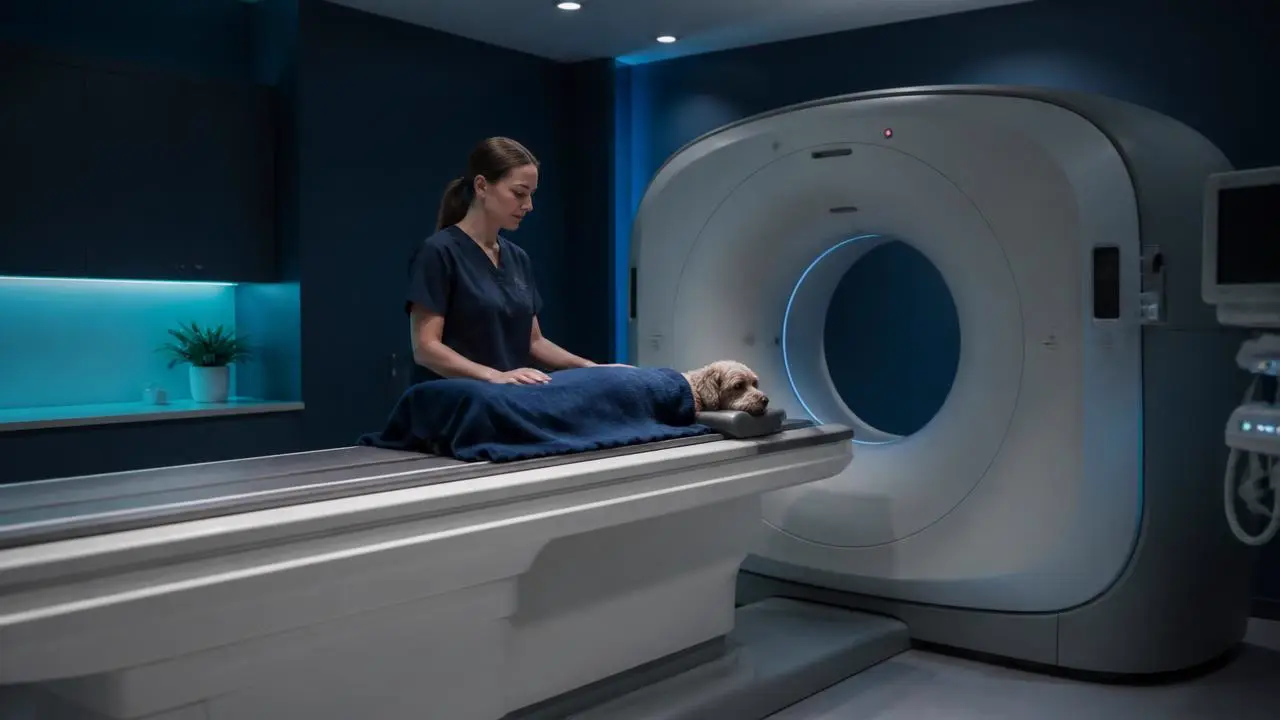
The cost of a CT scan for dogs is the single most common reason a study that would have changed treatment does not get taken. Not clinical disagreement, not availability: the number on the estimate. Some of that caution is entirely reasonable. Much of it rests on assumptions about veterinary CT cost that are either outdated or were never accurate, and the result is a diagnosis arrived at weeks late and often for more money in total. Here are the five that come up most.
By the RadsForVets editorial team, with clinical direction from Miriam M. Shanaman, VMD, MS, DACVR, Chief Medical Officer and board-certified veterinary radiologist. Published August 21, 2026.
Key takeaways
- The comparison that matters is not CT versus nothing, but CT versus the repeat-imaging and failed-treatment pathway that often precedes it anyway.
- Most of a CT fee is fixed infrastructure and the anaesthetic episode, not the scan time itself.
- Many insurance policies cover diagnostic imaging; the specialist report supports the claim.
- Modern scan times of under a minute have shortened the anaesthetic episode that drives most of the perceived risk.
- An unread or under-read CT study is where the real waste is: hundreds of images, no specialist impression.
Why cost myths delay diagnoses
A cost objection is rarely stated as one. It arrives as let us try treatment first, or let us repeat radiographs in three weeks, or let us see how she does. Those are sometimes the right call. They become the wrong call when the reason underneath them is an assumption about price that nobody has checked. Our pillar guide to veterinary CT scan interpretation covers the clinical side of when CT is indicated; this article deals with the money argument that sits in front of it.
Myth 1: CT is only for referral hospitals
The reality: scanner access has spread well beyond referral centres, and interpretation access has spread further still. A general practice does not need to own a scanner to use CT. Patients are commonly scanned at a nearby imaging facility or a collaborating hospital, and the resulting study is submitted for specialist interpretation remotely, with the report returning to the primary veterinarian who is actually managing the case.
The part that used to be genuinely restricted to referral centres was not the machine but the radiologist standing next to it. That constraint is gone. A two-doctor practice can obtain the same board-certified CT interpretation a teaching hospital gets, because the study travels instead of the specialist.
Myth 2: insurance never covers it, so there is no point asking
The reality: diagnostic imaging is covered under a great many policies, and CT is not usually singled out for exclusion. What varies is the tier of cover, the annual limit, and whether the condition is deemed pre-existing.
- Check before scheduling, not after. A five-minute call by the owner to confirm diagnostic imaging coverage and the remaining annual limit changes the conversation entirely.
- Submit the specialist report with the claim. A board-certified radiologist's report documents medical necessity in the terms insurers assess, far better than a procedure code alone.
- Pre-authorisation where offered. Some insurers will confirm coverage in advance, which removes the uncertainty that causes owners to decline.
Myth 3: CT is always more expensive than the alternative
The reality: it is more expensive than any single alternative step and frequently cheaper than the sequence of steps that replaces it. The honest accounting has to include everything the delayed pathway costs.
| Delayed pathway | Early CT pathway | |
|---|---|---|
| Imaging | Initial radiographs, then repeat series weeks later | Radiographs, then one CT study |
| Treatment | One or more empirical treatment trials | Targeted treatment from the start |
| Consultations | Multiple rechecks over weeks | Fewer, with a definitive answer earlier |
| Sedation episodes | Often several, for repeat imaging | Typically one |
| Time to diagnosis | Weeks to months | Same week |
| Total cost | Frequently higher once summed | Concentrated into one visible number |
The reason the delayed pathway feels cheaper is that its cost is distributed across several small invoices while CT arrives as one large one. That is a presentation problem, not an economic one. Our breakdown of what a teleradiology read actually costs per study makes the same point about interpretation fees specifically.
Myth 4: the sedation risk makes it not worth it for most patients
The reality: the anaesthetic episode is the real risk to weigh, and it has shrunk dramatically. Modern scanners acquire most studies in well under a minute, so what used to be a prolonged anaesthetic is now often light sedation with a short recovery.
- Scan time is not the same as table time. Positioning and monitoring dominate, and both are routine procedures for a trained team.
- Risk is patient-specific. A geriatric patient with cardiac disease warrants genuine caution. A stable middle-aged dog with chronic nasal discharge does not carry the same profile.
- Compare against the alternative. Repeat radiographic series over months often means several sedation events rather than one.
Myth 5: a scan without a specialist read is just as good
The reality: this is the myth that wastes the most money, because it spends the entire cost of the study and then declines the part that converts it into an answer. A three-view radiographic study is three images. A CT study is commonly several hundred, and a multi-phase contrast study can exceed a thousand, each needing review at more than one window setting because bone, soft tissue and lung are read from the same data differently.
- Systematic review of that volume is a trained discipline, not a matter of scrolling faster.
- Recognising normal cross-sectional anatomy across species and breeds is a separate skill from reading projections.
- A nodule invisible at soft tissue window is obvious at lung window; findings are missed at the display level before they are missed diagnostically.
- Reformatting into sagittal or dorsal planes is frequently what turns an ambiguous finding into a definite one.
- Higher sensitivity surfaces incidental findings, and deciding which warrant pursuit is judgment that prevents its own kind of harm.
Our article on what CT scans reveal that radiographs miss works through specific examples of findings that depend entirely on how the study is read.
What a veterinary CT scan actually involves
Separate from the cost argument, it is worth stating plainly what the study is, because owners and sometimes clinical teams picture something considerably more elaborate than the reality.
The study, step by step
- The patient is sedated or lightly anaesthetised, because any motion blurs the detail that justified choosing CT.
- They are positioned on the table, typically sternal or dorsal depending on the region of interest.
- The table passes through the gantry while the X-ray tube rotates around the patient. Acquisition takes seconds.
- If indicated, iodinated contrast is given intravenously and further sequences are acquired at timed phases.
- The computer reconstructs the projections into thin cross-sectional slices viewable in any plane.
- The dataset is transferred for interpretation and a report with findings, impression and recommendations comes back.
Recovery from the sedation is usually the longest part of the visit. For most patients the animal goes home the same day.
Free for veterinary teams
The CT cost conversation script
A short guide for the conversation with an owner who has just seen a CT estimate: how to frame total pathway cost, what to check with their insurer, and how to explain what the specialist report adds.
- Framing CT against the delayed pathway, not against nothing
- The insurance questions an owner should ask before scheduling
- Explaining sedation risk honestly and proportionately
- Why the interpretation is not the part to economise on
No contracts. No minimums. Radiologists on shift every day of the year.
Frequently asked questions
Sourced from real search questions people ask about this topic; answered by our team. These are drawn from Google autocomplete data for "cost of ct scan for dogs" and "veterinary ct scan". Answers are written by the RadsForVets editorial and clinical team, not derived from prompt-tracking data, which on this account currently returns mostly competitor-comparison and navigational queries rather than genuine consumer questions.
Cost and value
Why are dog CT scans so expensive?
Most of the cost is fixed infrastructure rather than the scan itself: the scanner, its service contract, the shielded room, the anaesthesia or sedation episode with a trained person monitoring it, and the specialist interpretation of several hundred images. A single facility spreads those fixed costs over its annual caseload, which is why the same study can differ substantially in price between a high-volume referral centre and a practice that scans occasionally.
Are CT scans covered by pet insurance?
Frequently yes, when the policy covers diagnostics and the condition is not pre-existing or excluded. Coverage varies enormously between insurers and tiers, so the practical advice is to have the owner confirm diagnostic imaging coverage and any annual limit before scheduling, and to submit the radiologist's report with the claim, since a specialist interpretation documents medical necessity clearly.
How much does a veterinary CT scan typically cost?
Ranges quoted to owners vary widely by region, whether contrast and multiple phases are used, the length of the anaesthetic episode and whether specialist interpretation is included or billed separately. Rather than quoting a number that will be wrong somewhere, ask your imaging facility for an itemised estimate that separates the scan, the anaesthesia and the interpretation, so you can see what is actually being charged for.
Are CT scans for dogs worth the cost?
When the result changes management, almost always. The comparison that matters is not CT against nothing, but CT against the alternative pathway: repeat radiographs, an empirical treatment trial that fails, weeks of delay and then the scan anyway. Where CT is not worth it is when the clinical question can be answered by a cheaper modality, which is exactly what asking a radiologist beforehand establishes.
What drives the price difference between a CT scan and an X-ray?
Three things. Equipment cost and maintenance are an order of magnitude apart. CT nearly always requires sedation or general anaesthesia with monitoring, while many radiographic studies do not. And interpretation workload differs enormously: a three-view radiographic study is three images, while a CT study is commonly several hundred reviewed at multiple window settings.
Safety and process
Are CT scans safe for dogs?
Yes, for the great majority of patients. CT uses ionising radiation, but a diagnostic study is a single short exposure event and the risk is negligible against the benefit of an accurate diagnosis. The more relevant risk is the sedation or anaesthesia, which modern scan times of well under a minute have made substantially shorter and safer than it was a decade ago.
How are CT scans done on dogs?
The patient is sedated or anaesthetised so there is no motion, positioned on the scanner table, usually in sternal or dorsal recumbency depending on the region, and passed through the gantry while the tube rotates. Acquisition itself takes seconds. If contrast is needed it is given intravenously and further sequences are acquired at timed phases. Total table time is usually far shorter than the recovery.
Do cats and exotic companion species need sedation for a CT scan?
Nearly always, yes, and for the same reason as dogs: motion blur destroys the fine detail that justifies choosing CT. Smaller patients also require thinner slices relative to body size to preserve resolution, so stillness matters more, not less. Protocols and anaesthetic planning are adapted to the species and the patient's condition.
What does a CT scan show that an X-ray cannot?
Anything obscured by superimposition, which is a great deal. Non-displaced fracture lines in complex regions such as the pelvis or tarsus, small pulmonary nodules near the heart or diaphragm, nasal turbinate and tympanic bulla detail, the relationship of an abdominal mass to its blood supply, and vascular anomalies visible only in a correctly timed contrast phase.
What is a CT scan for animals, technically?
A series of X-ray projections acquired around the patient and reconstructed by computer into thin cross-sectional slices, which can then be viewed in any plane. The underlying physics is the same as radiography; the difference is that the output is a volume of data rather than one flattened shadow image.
Working with RadsForVets
Why would a dog or cat need a CT scan?
The common indications are chronic nasal discharge, chronic ear disease, suspected occult or articular fractures, metastasis screening before major surgery, complex abdominal masses, portosystemic shunt mapping and multi-region trauma. In each of those, the finding or its absence directly changes what happens next.
Does RadsForVets read contrast-enhanced and multi-phase CT studies?
Yes. Contrast timing frequently determines whether a vascular anomaly is seen at all, so we read multi-phase studies routinely and will advise on phase timing before the scan if you ask. Protocol advice ahead of acquisition is part of the service, not a separate consultation.
How fast can a CT study be read on a STAT basis?
Our STAT target is about one hour from submission, and it applies to CT as well as radiographs. Routine CT studies come back within 24 hours. Radiologists are on shift 24 hours a day, every day of the year, so an overnight emergency scan does not wait for the morning.
Can a general practice submit CT studies without an in-house PACS?
Yes. We accept studies from practices that have no PACS of their own and will walk your team through submission from your existing equipment during setup. If your CT is done at a nearby imaging facility rather than in house, the study can still be submitted to us for interpretation.
Which species does RadsForVets read CT studies for?
Dogs, cats and exotic companion species. Our radiologists read small animal caseloads exclusively, which is what allows protocol advice to be specific rather than general.
Do I need a contract or minimum case volume for CT reads specifically?
No. The same terms apply to every modality: no contract, no sign-up fee, no minimum case count, and payment per completed case. A practice that sends one CT study a quarter is treated no differently from one that sends several a week.
Related reading
- Veterinary CT scan interpretationProtocols, reporting and when CT is the right modality over MRI or ultrasound.
- What nobody tells you about veterinary radiology servicesWhat sits inside the fee for a read, and what does not.
- Turnaround time versus accuracyWhy a fast CT read and a careful one are not opposites.
Work with us
Board-certified reads, 1 hour STAT, every day of the year
No contracts, no minimums, and direct access to the radiologist reading your case.